Cardiac arrhythmias – in a nutshell
Cardiac arrhythmias are irregular heartbeats that deviate from the normal course. Affected people sometimes perceive this as stumbling or racing of the heart. Is it dangerous? That is a big question. Thus the length of this article.
Our goal is to give you a framework to understand the different types of cardiac arrhythmias. They are mostly harmless, but can also be life-threatening.
Cardiac arrhythmias should therefore always be clarified by a doctor. An electrocardiogram (EKG) at rest, supplemented by a 24-hour long-term EKG and a stress EKG as well as an ultrasound examination of the heart help to track down the arrhythmia. Further examinations may also be necessary. If treatment is needed, it will depend on the type of abnormal heart rhythm.
What are arrhythmias?
The heart normally beats about 60 to 80 times per minute under rest conditions. The electrical activity that triggers the contraction of the heart muscle is generated in the heart itself: the clock is the so-called sinus node, which is located in the upper area of the right atrium. From here the impulses reach the AV node (atrio-ventricular node) via the walls of the atria. This structure usually represents the only electrical connection between the atrium and the ventricle. The AV node works to a certain extent as a “gatekeeper” with the combined properties of a conduction delay and a quick and orderly transmission of the electrical excitation via specific conduction pathways (His bundle, fascicle in the right and left ventricles and Purkinje fibers) into the muscles of the heart.
When you get excited or do some physical exertion, your pulse accelerates, while it slows down during sleep, for example. These changes are caused by the so-called autonomic nervous system, which influences both the sinus node and the AV node.
An irregular heartbeat sequence is called arrhythmia. Mild or occasional cardiac arrhythmias are often not noticed at all. The irregular heartbeat can also be felt as a “heart stumble” or a racing heart. It can lead to dizziness, nausea, restlessness, fainting, unconsciousness, seizures as well as chest pain and tightness, in the worst case even sudden cardiac death.
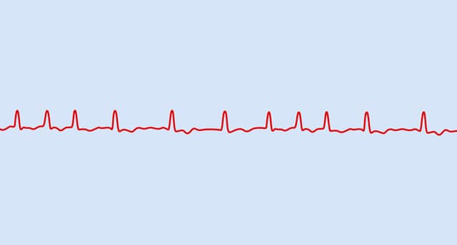
Using the heart current curve in the electrocardiogram (EKG), the doctor can recognize whether the heart has changed its normal, so-called sinus rhythm, into an irregular (arrhythmic) and/or too fast (tachycardia) or too slow (bradycardia) rhythm. It is important to find out the probable cause of the arrhythmia and, if possible, to correct it.
Causes
External causes of cardiac arrhythmias can be, for example:
• Nervousness, excitement and fear
• Excessive consumption of caffeine (for example in the form of coffee, energy drinks or cola)
• Alcohol consumption
• Consumption of drugs and poisons
• Side effects of some medications (for example thyroid hormones or certain psychotropic drugs)
• Severe bloating (meteorism)
• Febrile infections
• Irritation of the so-called carotid sinus node: This is a receptor on the main artery in the neck, which can be irritated, for example, by a tight scarf or collar, overstretching the head or blow/pressure. The result is a sharp slowdown in the heartbeat, leading to fainting. If the carotid sinus is oversensitive, it is called the carotid sinus syndrome.
Organic causes of arrhythmias include:
• Coronary artery disease (CHD)
• Myocardial infarction
• Myocardial diseases (cardiomyopathies)
• Inflammation of the myocardium (myocarditis)
• Heart valve defects
• Congenital diseases that lead to arrhythmias (for example Brugada syndrome, arrhythmogenic right ventricular disease, Wolff Parkinson’s disease -White syndrome, WPW syndrome )
• rarely high blood pressure (hypertension)
• electrolyte
• disorders (for example potassium deficiency)
• thyroid over- or underactive (hyperthyroidism, hypothyroidism)
Forms of arrhythmia
There are many forms of cardiac arrhythmias: A distinction is made on the one hand with irritation disorders (impaired generation of electrical impulses) and disorders with faulty transmission of the heart’s excitation (conduction disorders). Arrhythmias are also divided according to their place of origin (atrium or ventricle).
Furthermore, doctors differentiate between arrhythmias with a heartbeat that is too slow (called bradycardia and bradyarrhythmias; pulse at rest below 60 beats per minute) from those with too fast a heartbeat (tachycardia or tachyarrhythmias ; pulse at rest above 90 beats per minute).
Examples of cardiac arrhythmias are:
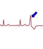
An extrasystole on the EKG
Extrasystoles: These extra beats can originate either from the atria (supraventricular extrasystoles) or from the ventricles (ventricular extrasystoles). Extrasystoles are usually not pathological. They occur in small numbers in almost everyone. Treatment may only be necessary if the extrasystoles exceed a certain level and the patient has symptoms, but especially if there is evidence of structural heart disease (e.g. scars from a heart attack, myocarditis, heart valve defects, diseases of the heart muscle).
Supraventricular tachycardia: rapid heartbeat, based on impulses in the atrium, either as a focal point of disturbance, for example due to previous damage to the heart, or as a circulating short-circuit excitation.
WPW syndrome: Congenital additional conduction between the atrium and the ventricle in the valve plane, which can lead to palpitations (tachycardia) due to extrasystoles. Since, in contrast to the AV node, this additional electrical connection has no line delay and the excitation in the ventricle does not run via the specific conduction system, indications of the underlying disorder due to the change in the premature ventricular excitation can often be seen in the resting ECG.
AV node reentry tachycardia: Second supply line in the AV node, which can also lead to a racing heart
Atrial fibrillation, atrial flutter: Rapid, irregular excitation in the atria, which leads to an irregular pulse. Due to the high, mostly undirected frequencies in the atria (approx. 400 beats per minute), these no longer contract, but only flicker. The ventricles and thus the heart beat irregularly and depending on the properties of the “gatekeeper structure” AV nodes are often too fast (more than 100 beats per minute), sometimes too slowly (less than 60 beats per minute). This affects the blood transport to the heart chambers, the heart pumps out too little blood.
Particularly in the left atrial appendage, a protrusion of the left atrium, the blood flow may even stall so much that clots can form. If these leave the heart via the large artery, they can reach the brain and trigger a stroke there.
If there is a structural change in the atria, the unphysiological, rapid atrial excitation (250-350 beats/min) can also take place in more orderly channels, such as with atrial flutter. Then there is a regular racing heart. The “gatekeeper structure” also protects against life-threatening heart rates and only transmits a fraction of the atrial actions – often in a ratio of 2: 1 to 4: 1.
SA block: Delayed or blocked transmission of excitation between the sinus node and the atrium (atrium).
Sick sinus syndrome: Slow heartbeat, sometimes alternating between tachycardia and bradycardia due to a malfunction of the sinus node.
AV block: A delayed (1st degree) or partially to completely blocked (II and III degree) transmission of the excitation between the atrium and the ventricle due to a disease of the AV node. If the blockage is higher, the heartbeat slows down. If the transmission is completely interrupted, a cardiac arrest can occur. A pacemaker is necessary to prevent this.
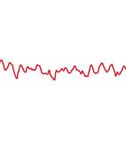
Ventricular fibrillation: dangerous for life
Ventricular tachycardia: racing heart due to additional pulses in the heart chamber. Ventricular tachycardias must be taken seriously – although in certain cases they can be “benign”, in other cases they turn into a life-threatening ventricular flutter or ventricular fibrillation.
Ventricular fibrillation, ventricular flutter: Uncoordinated, rapid electrical actions and tremors instead of controlled contraction of the chamber (with ventricular fibrillation over 320 beats per minute). The problem with this is that the heart pumps out little or no blood, it stands still. Without treatment (defibrillation), ventricular fibrillation ends fatally after a few minutes.
Symptoms: How do you notice cardiac arrhythmias?
Possible symptoms of cardiac arrhythmias include:
- Uncomfortably noticeable heartbeat (palpitations)
- Racing heart (tachycardia)
- Dizziness, lightheadedness, confusion
- Fainting, brief loss of consciousness (syncope), seizures
- Chest pain and tightness (angina pectoris)
Dreaded complications from cardiac arrhythmias can include:
- Embolism (vascular occlusion due to blood clots that are carried on)
- Stroke (cerebral infarction, apoplexy)
- Heart attack (myocardial infarction)
- Worsening heart failure (heart failure)
- Sudden cardiac death
When to see the doctor
If you feel that your heartbeat is irregular, the heart stumbles or suddenly races without explanation, you should see a doctor, even if you are otherwise fine. The same applies if the heart rate is too low. The doctor can get to the bottom of the matter and determine whether it is a cardiac arrhythmia and whether it can be classified as dangerous or harmless.
In the case of serious symptoms such as loss of consciousness, seizures, chest pain or shortness of breath, ambulance service is required.
Diagnosis
ECG: The heart rate measurement provides information about arrhythmias
The doctor will first inquire about the symptoms and previous illnesses. This is followed by a physical examination. When listening to the heart with the stethoscope, the irregular heartbeat is often already perceptible; unless the arrhythmias are seizure or random. Heart rate and blood pressure are also measured.
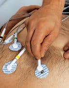
The most important test used to diagnose arrhythmias is electrocardiography (EKG). The electrical currents in the heart are measured via measuring points on the chest and arms and legs and recorded as a so-called electrocardiogram.
The ECG is initially performed under resting conditions (resting ECG). If necessary, a stress ECG (ergometry; measurement of cardiac activity under stress conditions, for example when running on a treadmill or cycling on a bicycle) can provide further information. Because certain cardiac arrhythmias only occur or worsen under stress. A long-term ECG over 24 hours or more helps to reveal irregularities in the heart rhythm that only occur sporadically. Another option is to give those affected a small portable EKG machine and ask them to start a recording if symptoms occur (event recorder). In this way, cardiac arrhythmias that rarely occur can possibly be recorded. If the doctor suspects cardiac arrhythmias as the cause of fainting attacks (syncope) or strokes, small devices implanted under the skin (loop recorders) are now able to monitor and store the heart rhythm for up to three years. In addition, some electrical watches and other so-called “wearables” now have the option of registering and saving an EKG and roughly assessing the findings. Of course, these cannot replace an analysis by the specialist.
The doctor often also does an ultrasound examination of the heart, a so-called echocardiography. Changes in the structure and movement of the heart, valve defects and other possible causes of cardiac arrhythmias are recorded.
In most cases, these examinations are sufficient to make the diagnosis of a cardiac arrhythmia. In the case of special questions, further tests, for example, heart current measurements with a cardiac catheter (known as electrophysiological examination or EPU) or examinations with the administration of certain medications, may be necessary.
Therapy
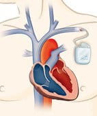
Sometimes the solution: a pacemaker
Cardioversion is the restoration of the normal heart rhythm. It can be done with medication or with the help of a defibrillator. This so-called electrocardioversion is used as an emergency treatment for ventricular flutter and ventricular fibrillation. A strong current surge first interrupts the electrical activities in the heart and thus enables a new beginning from the sinus node. For atrial flutter and atrial fibrillation, electrical cardioversion under short-term anesthesia is also an option if medication cannot normalize the heart’s activity.
Another option for treating cardiac arrhythmias is so-called ablation with heat (high-frequency ablation) or cold (cryoablation). The tissue that was identified as the starting point of the cardiac arrhythmia is desolated so that it no longer generates or conducts excitation. The treatment is carried out using special cardiac catheters and has proven itself in certain types of palpitations, such as AV node reentry tachycardia, atrial flutter and certain arrhythmias in the ventricles. Even if – as in WPW syndrome – there are additional conduction pathways between the atria and the ventricle, an ablation can be useful. In the case of atrial fibrillation, which has not been around for a long time, and cannot be eliminated by medication and which affects the patient’s condition and performance, cardiologists recommend catheter ablation by completely isolating the usually four pulmonary veins that open into the left atrium. Here the success rates with regard to the permanent prevention of a recurrence of atrial fibrillation are up to 80 percent.
If dizziness, fainting, shortness of breath and reduced performance are due to a pulse that is too slow or partially interrupted (total AV block or pathological sinus node), a pacemaker is necessary. Pacemakers are small, battery-operated devices that send electrical impulses to the heart and thus normalize the heart rhythm. They are placed near the heart, under the collarbone, in a small surgical procedure. The pacemaker sends its impulses to the heart via thin probes that are inserted into the superior vena cava, the right atrium and/or the right ventricle. Mini pacemakers without probes are new if only right ventricle stimulation is required. These are small wireless devices (cardio capsules) that are anchored in the right ventricle of the heart.
In the case of severe pumping weakness in the left ventricle (for example after a heart attack or after an inflammation of the myocardium) or in the case of certain hereditary heart diseases, sudden cardiac death from ventricular fibrillation can be prevented by implanting a defibrillator (ICD) in the patient. It generates a surge of electricity that is supposed to restart the heart’s action. Alternatively, there are ICDs in which the probe(s) are not implanted into the heart via a vein, but are instead guided under the skin parallel to the heart along the sternum (subcutaneous ICD). If only temporary protection against ventricular fibrillation is likely to be necessary, an external defibrillator such as a vest can now be worn for up to three months.
Self-help

What you can do against cardiac arrhythmias:
- Avoid stress and excitement – try to relax more often and give yourself enough rest.
- If you suffer from cardiac arrhythmias, you should avoid excessive consumption of caffeine and alcohol (alcohol is particularly bad for atrial fibrillation!).
- No Smoking – if necessary try to get help with hypnosis or in an addiction clinic.
- Are you taking any medicines? Talk to your doctor about whether these can lead to palpitations. A change in preparation or a different dosage may be necessary.
- Go to your doctor for regular preventive care. Disorders of other organs can also be behind cardiac arrhythmias, for example, an overactive thyroid.
- If you have a pacemaker or an ICD, it is essential that you keep your appointments for a pacemaker check-up! If problems arise in between, see a doctor immediately!
Of course, colon hydrotherapy can’t fix your heart problems, but doing a blood and brain booster certainly can’t hurt.


0 Comments